Bovine Adenovirus (BAdV) in R1 Dairy Calves

HISTORY

We recently had a case of BAdV confirmed in a mob of 140 rising yearling dairy replacements. Five rising yearlings had died over a period of 3-4 days. Unfortunately, four of them had died over the previous 24-48 hours during a very warm spell of weather, and when found were too autolyzed to postmortem. The fifth calf, noticed late the next day, was acutely unwell, and subsequently examined, treated and blood tested/faecal sampled. The calf had a high temperature, dull demeanour, dehydrated, diarrhoea and unable to stand unaided. This calf died overnight. The mob’s drenching history was up to date, and unlike a lot of reported cases of BAdV, where the mob has been unwell, the remaining calves appeared in ‘good health’.

THE DISEASE

BAdV is a relatively uncommon disease – but was noted as a disease of increasing occurrence in rising yearlings in a national review 10 years ago. It is a viral disease that is mainly seen in 6-12 months old calves. Outbreaks, usually with low mortality rates (1-3%) can occur during autumn, winter and spring months, and often post weaning. It is primarily an acute gastro-intestinal disease, but calves may also have respiratory signs.

Adenoviruses are transmitted in nasal and oral secretions and faeces. While many cattle are infected, only a small proportion develops disease, which are animals usually immuno-compromised or have concurrent BVD infection. Common differentials for BAdV are Yersinia, Salmonella, BVD/mucosal disease, GI parasites, and other causes of acute death – nitrate poisoning, clostridial disease or toxicity.

DIAGNOSIS

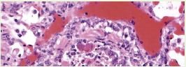
Until recently, the main way to diagnose BAdV, has been getting good quality bowel samples and doing histopathology – and seeing the classic inclusion bodies, of this disease. However, a PCR (looking at genetic material) blood test is now available – and markedly assists us with the diagnosis. The fifth calf was positive for BAdV, and it was assumed it was also the cause of the other four calves that died.

Balancing the Winter Ration

To ensure a successful outcome to cow wintering, consideration needs to be made around cow requirements for energy, protein and minerals (Ca, P and Na). Knowing the content of the diet, the proportions to be fed and the likely utilisation will enable you to fine tune where required. A dry cow needs a minimum of 10-11% crude protein and this increases to 16% as she nears calving.

Last year our practice did a large amount of feed testing on Fodder Beet crops. The range and variation in protein and macromineral content was enormous. A very low crude protein/calcium FB crop coupled with a low protein/calcium cereal/maize silage will not adequately support a pregnant cow or set her up for the following lactation (and reproductive season).

A large part of the protein and calcium in a fodder beet plant is held in the leaf. Although the leaf yield in most crops is looking good right now, as they exhaust soil of nitrogen and potassium reserves this may lead to leave senescence (die back of exterior leaves to pull reserves into the bulb). Consider the late applications of around 50 units of N and K/hectare to maintain the green leaf mass.

Look to get your winter diet tested now to provide time to make tweaks with supplement matches if these are required. Contact your Prime Vet about collection, testing and diet balancing.

Staph aureus is in 85% of herds – often causing minimal issues

The latest DairyNZ mastitis fact sheet states that about 85% of herds will have some cows that are infected with Staph aureus. It goes on to say that Staph aureus accounts for about 10% of clinical mastitis cases but can be significantly more in an out-break situation.

There are options for screening your herd to find the Staph aureus cows, and either cull them or manage them in such a way as they don’t spread the infection.

Remember though, there are many herds out there where there is Staph aureus found in the Bulk Milk (we find it regularly in the Antibiogram tests, even in low BMSCC herds). Staph aureus in the Bulk Milk is not causing a significant issue, so before you decide to test and cull your way to being “Staph free” decide … do you have an issue and, what are the herds that have Staph but not an issue doing that you aren’t.

What is happening with infected herds that don’t have an issue, knowingly or unknowingly, is the cows are managed and that infection is not being spread from cow to cow, so the percentage of infected cows is low.

The main source of Staph aureus is infected milk from infected quarters, being transferred on liners or milker’s hands to uninfected cows. Effective practices to slow or prevent the spread of Staph aureus are:

• Having clean hands – this really means wearing gloves, wet gloves are cleaner than wet hands.

• Using teat-spray on every teat at every milking will reduce the spread by 50%.

• Have teats that are not cracked or have teat end lesions – these teats are harder to disinfect, and teat end lesion suggests there is over milking occurring or the vacuum is excessive, or the pulsation is incorrect, or the liners are well past their life expectancy.

• These farms usually have good records so when they have a cow that gets a repeat mastitis case, before treating for the 3rd or 4th time, they consider drying the quarter off, drying off the cow early, or even culling.

If your farm set up allows it, consider running the 3 titters, high SCC cows and cows that have been treated for mastitis in a mob that gets milked last – or at least not in the first herd – so any contaminated liners end up on less cows. If you are considering testing the whole herd for Staph please phone your prime vet at the Veterinary Centre. There are numerous options out there, each with their pros and cons.

Yersinosis in Calves

Yersiniosis frequently rears its head in the months of December through to May in calves of 4-10 months of age. Calves with the disease suffer a chronic scour, elevated temperature, loss of appetite and weight loss. It is caused by the bacteria Yersinia pseudotuberculosis, which can be found in the gut in over 85% of herds of yearling heifers around the country and up to 60% of individuals. Although it is commonly found in the gut of healthy calves, an overgrowth of the bacteria and invasion of the gut lining results in millions of micro-abscesses throughout the length of the intestines.

Conditions which may precipitate an overgrowth in the gut are low levels of the trace elements copper and selenium, parasitism, BVD, long transport, poor feed quality or feed stress.

Once an outbreak starts, an insidious spread through a mob of calves occurs via the faecal-oral route.

Control and treatment comprise the removal of affected animals and use of appropriate antibiotic. Further prevention should focus on the underlying risk factors – correction of trace mineral status, drenching and increasing feed allocation. We have seen a lot of success with the use of Multimin injection.

There is a registered Yersinia vaccine available for deer. This has been used off-label in cattle but can result in anaphylactic reaction. We have a limited number of clients that have used a single dose of this vaccine with apparent protection inferred. You would need to talk with your prime vet about the pros and cons of using this product off-label.

Correctly Identifying the Lame Claw

Over the years I have witnessed multiple occasions where farm staff have attacked hooves with knives, grinders and hoof trimmers before correctly determining the site of lameness. This will have frequently resulted in an exacerbation of the overall lameness in the cow.

Before putting any lame cows into the crush, it is important to determine which leg is affected. Our vets tend to record these on a recording sheet. Look for:

  • obvious swelling (and hoof shape)

  • weight bearing while standing still (the lame foot will be positioned so not to carry as much load)

  • a head nod while walking in the case of front leg lameness

  • the stride length and speed (the lame leg will have a short stride before taking weight, then the sound leg swings through with a longer stride and is weight loaded for longer.

Once you have determined the lame leg, have tied it up, washed it down and checked for footrot, use hoof testers to confirm where the lameness is located. This simple tool works by squeezing and putting pressure on the claw. Watch for a repeatable pain response which is easy to recognise in the hind legs by a tensing of the ‘hamstring’ muscles and the front legs by a tensing of the shoulders. Cows with sole bruising will often have very soft hooves and you will see that the testers easily depress the sole. If there is no response to either claw to hoof testers, then chances are you either have the wrong foot or the cause of lameness is located further up the leg.